02.02 Early cells and microscopes

Module 02: Life's Origins

The big idea Cells are the basic units of life. Their structures distinguish cell types, and the microscope you choose determines which details you can see.

By the end you can

  • What is made of cells?
  • The three parts of cell theory
  • Scientists who built the evidence
  • Prokaryotes and eukaryotes
  • What both cell types share
  • The bacterial model
  • Magnification and resolution
  • Dissecting and compound light microscopes
  • TEM and SEM
  • Practice the virtual microscopy task
Build the idea in small steps

Cells are the basic units of life. Their structures distinguish cell types, and the microscope you choose determines which details you can see.

Not sure about the basics underneath this lesson?

What is made of cells?

All living organisms consist of one or more cells. Atoms build molecules; molecules build cell structures. A molecule such as water, DNA, or protein is not itself a cell. Milk and saliva are products of cells. Viruses are not cells and are treated as nonliving in this course.

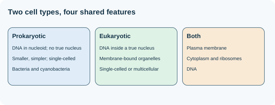
Both cell types have DNA, cytoplasm, ribosomes, and a cell membrane.

The three parts of cell theory

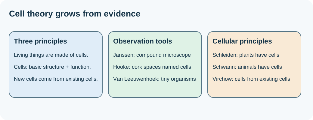
All living things are made of cells. Cells are the basic units of structure and function. New cells come from existing cells. The theory developed through observations, experiments, and contributions from many scientists; a scientific theory is an explanation supported by evidence.

Observations and improved instruments helped build the theory.

Scientists who built the evidence

The course credits Janssen with the early compound microscope. Hooke observed cork and named its boxlike spaces cells. Van Leeuwenhoek observed tiny living organisms. Schleiden concluded plants are made of cells; Schwann concluded animals are made of cells. Virchow supported the principle that cells come from preexisting cells. Redi and Pasteur helped disprove spontaneous generation.

Match each scientist to the observation or principle.

Prokaryotes and eukaryotes

Prokaryotes are generally smaller, simpler, and single-celled. Their DNA occupies a nucleoid region without a surrounding nuclear membrane; they lack a true nucleus and membrane-bound organelles. Bacteria and cyanobacteria are examples. Eukaryotes have a membrane-enclosed nucleus and internal membrane-bound organelles. Plants, animals, fungi, and protists are eukaryotes; some eukaryotes are single-celled.

Presence of a true nucleus is the key distinction.

What both cell types share

Both have a plasma membrane, cytoplasm, DNA, and ribosomes that make proteins. A cell wall supports and protects many cells, including the bacterial and plant models in the lesson; animal cells have a membrane but no cell wall. Membranes regulate exchange, and specialized structures help organisms survive in their environments.

A membrane belongs in both columns of a cell comparison.

The bacterial model

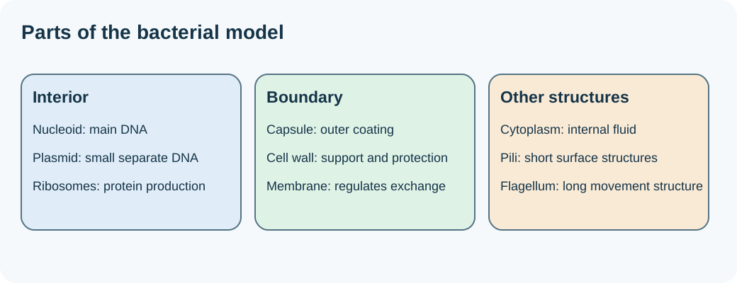
The nucleoid contains the main DNA. Ribosomes make proteins. Cytoplasm is the internal fluid, and the plasma membrane controls exchange. The wall supports and protects the cell. The notebook also labels a capsule, pili, plasmids, and a bacterial flagellum. A capsule is an outer coating; pili are short surface structures; plasmids are small separate DNA molecules; a flagellum helps movement.

Compare the bacterial structures named in the notebook.

Magnification and resolution

Magnification describes how much larger an image appears. Resolution describes how clearly you can distinguish close details. Making a blurry image larger does not supply new detail. Light microscopes use light and glass lenses; electron microscopes use electron beams and electromagnets and can resolve much smaller structures.

Choose the instrument for the detail you need.

Dissecting and compound light microscopes

A dissecting microscope uses reflected light from above to examine a specimen's surface at relatively low power, up to about 40Γ— in the lesson. It suits leaves, organs, and visible small objects. A compound light microscope shines light through a thin specimen from below. It suits cells, blood, and pond microorganisms and usually offers 40Γ— to 400Γ— or 1,000Γ— magnification. Light microscopes can be used with living specimens.

Whole surfaces versus thin, light-transmitting specimens.

TEM and SEM

A transmission electron microscope (TEM) sends electrons through a thin specimen to reveal internal details. A scanning electron microscope (SEM) scans the surface and gives a detailed view of surface shape. Electron microscopes have higher resolving power than light microscopes, but specimen preparation prevents viewing living cells. Images begin in black and white; added color is artificial.

TEM: through the specimen. SEM: surface.

Practice the virtual microscopy task
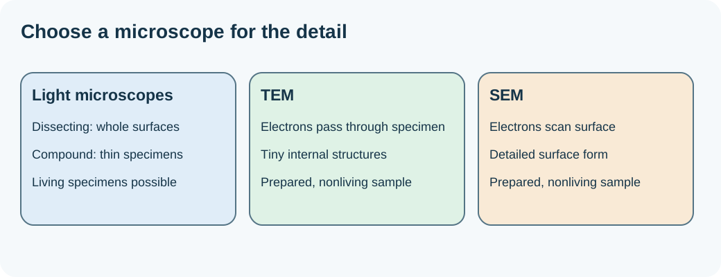
Compare what each instrument reveals, how it works, and its limitations. For living pond microorganisms, choose a compound light microscope. For a whole leaf's surface at low power, choose dissecting. For tiny internal details, choose TEM. For detailed surface form, choose SEM. Record actual observations in your course notebook when completing the assigned activity.

Explain your choice using both the specimen and the needed detail.