Module 03 — Coordination and Control · quiz worth 20 pts · 2026-10-21
The big ideaAn action potential is a moving change in membrane voltage; at a neuromuscular junction, it triggers acetylcholine release and starts a muscle-fiber action potential.
Keep three locations separate
Most mistakes mix events in the axon membrane, terminal, and muscle membrane.
Acetylcholine receptors create an end-plate potential that can start a muscle action potential.
Quick check — answer out loud before you peek
Which ion enters the motor-neuron terminal to trigger vesicle release?
Calcium.
Got it now? Scroll back up and re-read the section — it should land differently.
Resting voltage stores a signaling possibility
Sensory neurons, interneurons, and motor neurons are the basic building blocks of the nervous system. At rest, the inside of a neuron is negative and the outside is positive. That difference across the membrane is the membrane potential; at rest it is called the resting potential. Protein channels and sodium-potassium pumps maintain it: the pump keeps the ion gradients in place over time, while gated channels create the rapid changes of an impulse. A nerve impulse (action potential) is a reversal and restoration of that charge. It moves in one direction and can travel up to 100 meters in less than a second.
Threshold triggers an all-or-none event
A stimulus that reaches threshold opens sodium gates. Sodium rushes in and the membrane depolarizes: the inside switches from negative to positive. Each depolarized section pushes the next section to threshold, like falling dominoes. At the peak, sodium gates close and potassium gates open; potassium streams out and the membrane repolarizes. The lesson puts the time between depolarization and repolarization at about 3 milliseconds.
Read the action-potential graph
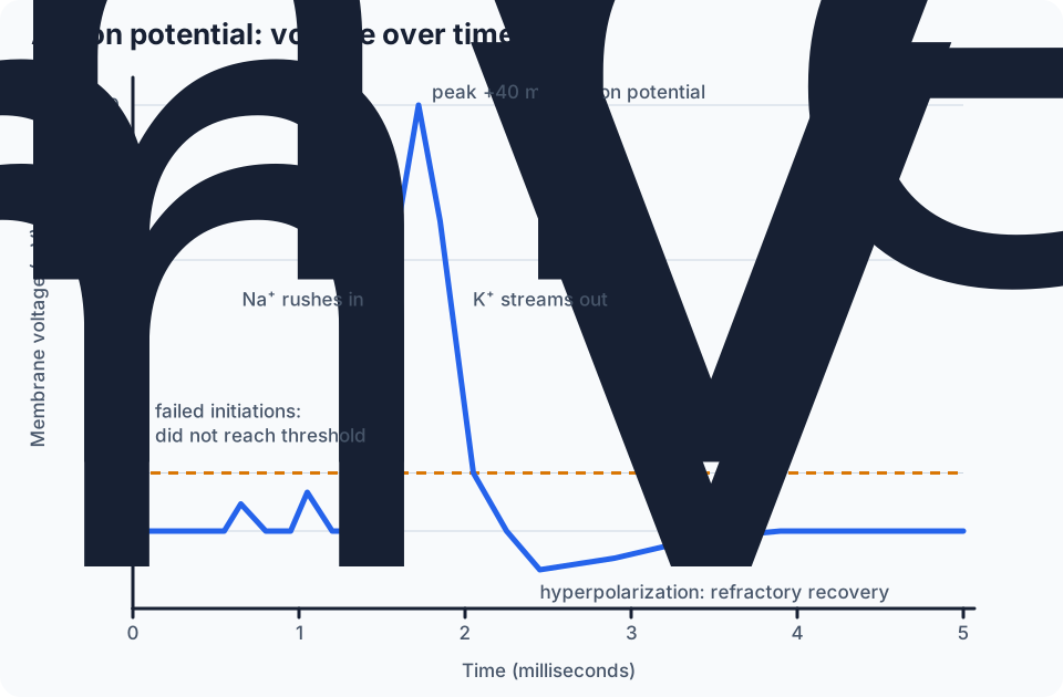
Read the graph left to right: flat resting potential at −70 mV; two failed initiations that do not reach the −55 mV threshold; a steep depolarization to +40 mV; a steep repolarization; a dip below rest (hyperpolarization), which the course graph labels the refractory period; then a slow return to rest. Refractoriness actually begins during the spike itself and continues through that dip.
The peak is the electrical impulse. It stimulates an action potential in the next region of the neuron, moving the impulse along until it reaches the end of the axon.
Rest −70 mV, threshold −55 mV, peak +40 mV. Only the stimulus that reaches threshold makes an action potential.
Not sure about this bit?
Use the three numbers on the voltage axis.
1
−70 mV
Flat line here is rest. Below it after the spike is the refractory period.
2
−55 mV
Threshold. Bumps that stop under it are failed initiations.
3
+40 mV
The peak. The climb is depolarization; the fall is repolarization.
Got it now? Scroll back up and re-read the section — it should land differently.
The refractory period supports one-way travel
From the start of the spike through the below-rest dip, that section of membrane cannot fire normally again. The next section is brought to threshold while the previous one recovers, so the impulse keeps moving forward.
Synapses and neurotransmitters
Most neurons do not touch one another. The space between two neurons is a synapse; the space between a neuron and a muscle cell is a neuromuscular (myoneural) junction. A neurotransmitter carries the impulse across like a bridge. Dopamine and norepinephrine are neurotransmitters that help control coordination and muscle movement, heart rate, circulation, and metabolism.
Substances can change synaptic transmission. In the lesson, caffeine increases it, which can cause nervousness and sleeplessness; LSD, mescaline, and amphetamines interfere with how synapses work; and MDMA disrupts neurons that use serotonin, a neurotransmitter that regulates mood, sleep, and sensitivity to pain.
The axon terminal changes electrical to chemical
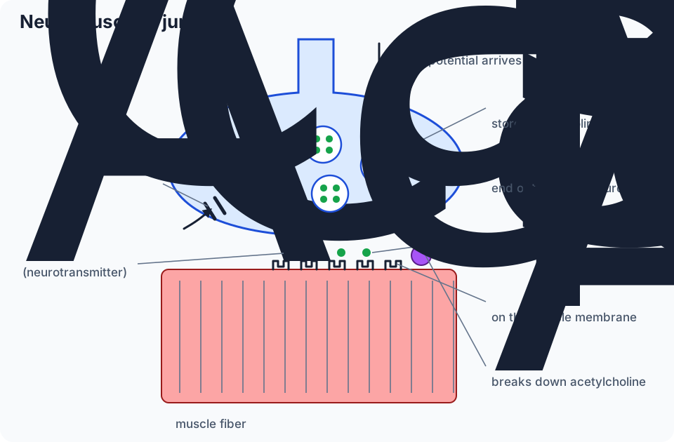
When the motor neuron's action potential reaches the axon terminal, a calcium channel lets calcium in, which signals the synaptic vesicles to release acetylcholine into the synaptic cleft. Each vesicle stores about ten thousand acetylcholine molecules. Acetylcholine diffuses across the cleft to motor end plate receptors on the muscle membrane.
Six structures to know: axon terminal, calcium channel, synaptic vesicle, neurotransmitter, synaptic cleft, motor end plate receptor.
Not sure about this bit?
Six labels, from the top of the drawing down.
1
Nerve side
Axon terminal, its calcium channel, and the synaptic vesicles inside it.
2
The gap
Synaptic cleft, with neurotransmitter (acetylcholine) crossing it.
3
Muscle side
Motor end plate receptors on the muscle membrane.
Got it now? Scroll back up and re-read the section — it should land differently.
The muscle changes chemical back to electrical
Acetylcholine briefly bonds to the receptor protein in the motor end plate. That opens the sodium and potassium gates of that region at the same time and creates an end-plate potential. Instead of firing there, the end-plate potential stimulates neighboring regions of the muscle to make true action potentials, which excite contraction. This link between the muscle's action potential and contraction is excitation-contraction coupling. Then acetylcholinesterase deactivates acetylcholine as soon as it detaches, so no extra contraction starts.
What the histology slides show
The lesson's seven microscope slides are not stored in the family library, so look at them in the course lesson itself. Use the captions to know what to look for.
Slides 1–3 (neuromuscular junction at 10X, 20X, 40X): efferent nerve fibers meeting skeletal muscle cells. Skeletal muscle cells are electrically excitable and controlled by neurons whose cell bodies are in the ventral horn of the spinal cord.
Slides 4–7 (axons and axon terminals at 4X to 40X): axons ending beside muscle fibers. Some axons carry sensory information to the CNS; others carry motor messages to skeletal muscle. The 40X slide labels the axon and the axon terminal, where acetylcholine is released.
Practice the voltage-to-movement chain
Separate ion movement in the axon from neurotransmitter movement across the junction. Some questions use numbered diagrams.