Module 03 — Coordination and Control · quiz worth 18 pts · 2026-10-20
The big ideaPeripheral pathways carry sensory input and motor output; somatic reflexes and autonomic circuits organize fast responses without requiring conscious control.
Put every PNS question on an arrow
Direction and target distinguish sensory, somatic motor, and autonomic pathways.
Not sure about the basics underneath this lesson?
1
Toward CNS
That is sensory or afferent input.
2
Toward skeletal muscle
That is somatic motor output.
3
Toward smooth/cardiac muscle or gland
That is autonomic output, usually through a ganglion.
Quick check — answer out loud before you peek
In an autonomic path, which neuron runs from a ganglion to the organ?
The postganglionic neuron.
Got it now? Scroll back up and re-read the section — it should land differently.
The PNS links receptors, CNS, and effectors
The peripheral nervous system is made up of the nerves that extend beyond the brain and spinal cord. It is divided into the somatic and autonomic nervous systems. Receptors detect a change; effectors respond to it. Afferent (sensory) neurons carry signals toward the spinal cord. Efferent (motor) neurons carry signals out to muscles.
Neurons from CNS to effector. Somatic: One; Autonomic: Two
Effect on muscle. Somatic: Excites; Autonomic: Excites or inhibits
Simple receptors and complex receptors
The somatic system is made up of some cranial nerves and all spinal nerves, and its nerves contain both sensory and motor neurons. Receptors in the skin, eyes, mouth, nose, ears, and muscles detect the change first.
Simple receptors are fairly small and spread throughout the body; they handle information such as a temperature change on your skin. Complex receptors serve the special senses: taste, smell, hearing, balance, and vision. Each sensory organ is built to receive one kind of stimulus well.
The special senses route a stimulus to the brain
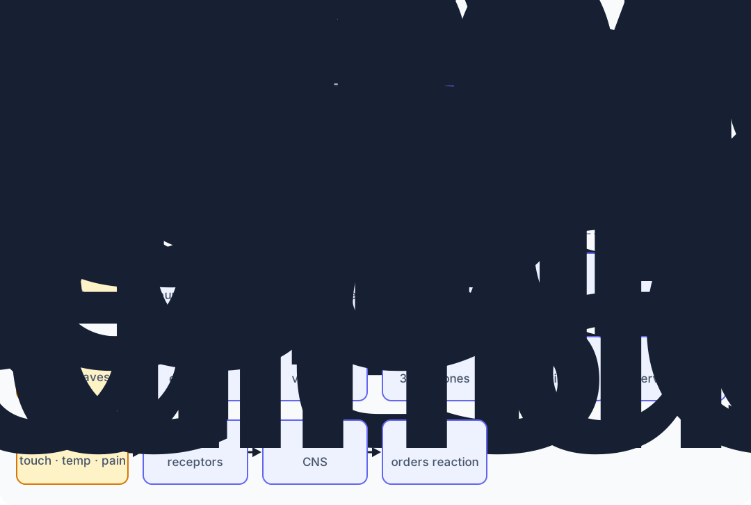
Smell: vapors enter the nasal cavity, which is lined with millions of olfactory sensors. These nerve cells turn the chemical message into a nerve impulse that travels to the olfactory bulbs, which send it to the brain.Taste: papillae on the tongue hold bundles of taste receptor cells called taste buds. The lesson video lists four basic flavors (sweet, salty, sour, bitter); current descriptions add umami (savory) as a fifth. The nose adds the finishing touches to flavor.Vision: the iris contracts and relaxes to control how much light enters. The lens focuses an upside-down image on the retina, the retina converts light to nerve impulses, the optic nerve carries them, and the brain turns the image right-side up.Hearing: sound passes the pinna and ear canal to the eardrum, which vibrates. The vibration crosses the three ossicles (the tiniest bones in the body) to the cochlea, whose cilia vibrate and stimulate the cochlear nerve.Skin: tactile nerve receptors sense pressure, vibration, temperature, and pain. The CNS analyzes that information and orders a reaction.
Each row follows the lesson's Senses video from stimulus to the brain or CNS.
Not sure about this bit?
Every sense follows the same three-step shape.
1
Receptor
A specialized organ part detects one kind of stimulus: olfactory sensors, taste buds, retina, cochlea cilia, or skin receptors.
2
Impulse
The receptor turns that stimulus into a nerve impulse.
3
Brain or CNS
A nerve carries the impulse in to be interpreted, such as the optic or cochlear nerve.
Got it now? Scroll back up and re-read the section — it should land differently.
A reflex arc favors speed
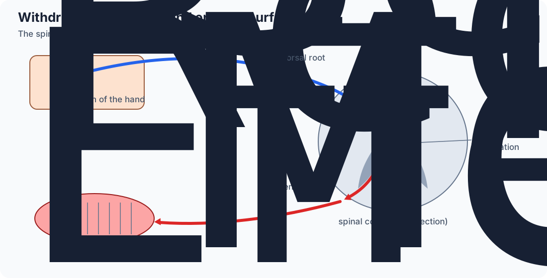
A reflex is an involuntary response to an internal or external stimulus. It is the one involuntary movement the somatic system controls. In the withdrawal reflex, the spinal cord, not the brain, processes the “my hand is on fire” signal and sends the “pull it away” response, which saves time. The signal also travels on to the brain, so you feel the pain, but the withdrawal does not wait for the brain to decide.
The withdrawal reflex uses five parts, in order: the receptor picks up the stimulus; the sensory neuron carries the impulse through the dorsal root into the spinal cord; an interneuron connects the afferent and efferent neurons; the motor neuron leaves through the ventral root; the effector (a muscle, gland, or organ) responds.
The knee-jerk is different. Tapping the patellar tendon stretches the quadriceps, and stretch receptors (muscle spindles) in the quadriceps send a sensory signal to the spinal cord. There the sensory neuron connects directly to the motor neuron for the quadriceps, with no interneuron in that link (a monosynaptic reflex). The quadriceps contracts and the lower leg kicks forward. The course video traces the signal from the patellar tendon and uses the five-part arc for every reflex; the five-part chain fits the withdrawal reflex.
Most reflexes are internal and unseen, such as digestion signals after you eat or a rise in blood pressure during exercise.
Withdrawal reflex: receptor → sensory neuron → interneuron → motor neuron → effector. The spinal cord starts the withdrawal; the brain also receives the signal and becomes aware of the pain.
The autonomic system controls involuntary effectors
The autonomic system controls cardiac muscle, smooth muscle, and glands. The sympathetic division prepares the body for an emergency (“fight or flight”). The parasympathetic division brings the body back to homeostasis.
Blood vessels. Sympathetic: Constrict to raise blood pressure; Parasympathetic: Dilate to lower blood pressure (course chart; most vessels are mainly under sympathetic control)
This is the course's simplified comparison. The first seven rows come from the lesson's comparison chart; mouth, liver, and bladder come from its nervous-system diagram, which the guided notes ask you to add.
Autonomic output uses a two-neuron chain
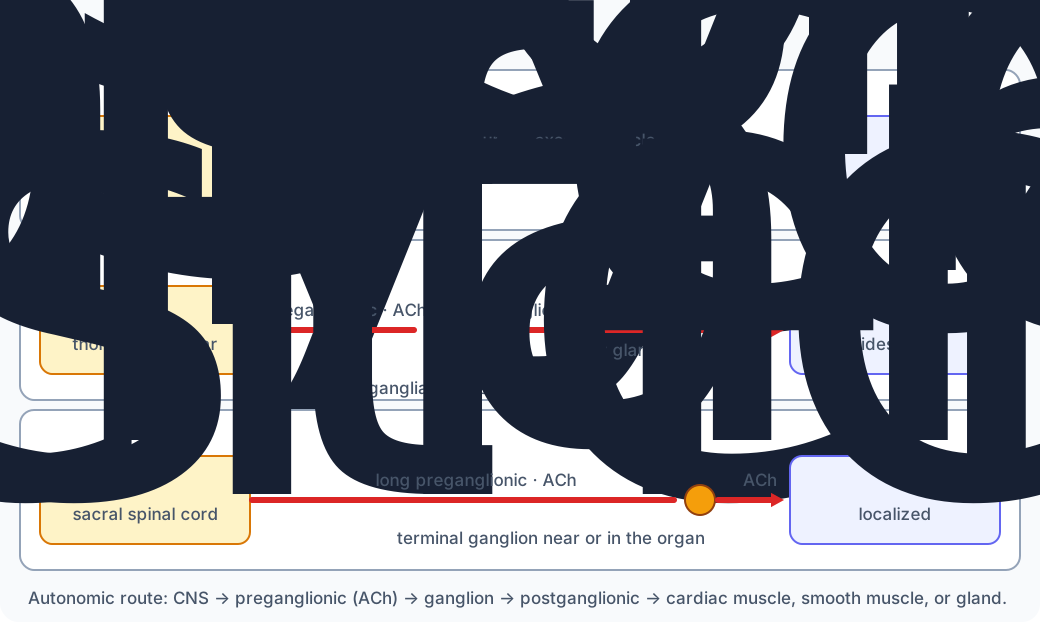
Ganglia are clusters of cell bodies in the PNS. A preganglionic motor neuron sits in the CNS and sends its fiber into a ganglion. A postganglionic motor neuron runs from the ganglion to the body organ. In both divisions, the preganglionic neuron releases acetylcholine at the ganglion.
Sympathetic: starts in the thoracic and lumbar spinal cord. Many of its ganglia form a chain just outside the spinal cord, which lets a strong emergency signal reach many organs together (the course calls this a whole-body response). Most of its postganglionic neurons release norepinephrine; those to sweat glands release acetylcholine instead.Parasympathetic: starts in nuclei of the brain stem, whose fibers leave in cranial nerves such as the vagus nerve (to the heart, lungs, digestive tract, and liver), and in the sacral spinal cord. Its ganglia are terminal ganglia near or within the target organ, so its effects are localized to specific organs. Its postganglionic neurons release acetylcholine.
A neurotransmitter's effect depends on the receptor and target tissue, not on the chemical alone. Norepinephrine can excite or inhibit; acetylcholine stimulates digestion but slows the heart.
Wording note: the course lesson describes parasympathetic ganglia as grouped along cranial nerves and calls norepinephrine the sympathetic transmitter without exceptions. If a course question uses that wording, it is pointing to the parasympathetic or sympathetic division; the placement and exception above are the anatomically accurate version.
Somatic output uses one motor neuron; autonomic output uses two, with a ganglion between them.
Not sure about this bit?
Compare the two divisions on four features.
1
Where it starts
Sympathetic: thoracic/lumbar cord. Parasympathetic: brain stem (cranial nerves such as the vagus) and sacral cord.
2
Ganglia
Sympathetic: a chain beside the cord. Parasympathetic: terminal ganglia near or in the target organ.
Got it now? Scroll back up and re-read the section — it should land differently.
Disorders of the peripheral nervous system
Autoimmune disorders, injury, or environmental factors can change the pathway the nerve signal travels.
Guillain-Barré syndrome is an autoimmune condition in which the body attacks nerves of the PNS, usually set off by an illness or stress to the body. Mild cases can bring severe pain or limited use of a body part; some cases make breathing difficult enough that the patient needs help.Diabetes is not itself a PNS disorder, but too much glucose in the blood can damage peripheral nerves, causing numbness or paralysis. Damage to peripheral blood vessels can mean blood no longer reaches the extremities, which can require amputation of toes or feet.
The lesson names these supports when the damage is known: treatments that help the nerve regrow or be stimulated, physical therapy, medications such as steroids that reduce inflammation, and devices that stimulate the nerve or block neurotransmitters to lower pain.
Practice the peripheral routes
Track direction, neuron count, and target. Some questions use numbered diagrams: name the numbered part.